
Facial contouring surgery does not mean that every part of the face should be made smaller.
A natural-looking facial contour is created by reducing only the areas that are excessively prominent and restoring volume only where it is genuinely lacking. Some patients want to soften an angular face but are concerned that facial contouring surgery may leave the face looking flat or hollow. In selected cases, a combined approach using 3-area facial contouring surgery—zygoma reduction, long-curve mandibular angle reduction, and T-shaped genioplasty—together with paranasal augmentation and autologous fat grafting can address both concerns.
Rather than focusing only on bone reduction, the surgical plan should also consider the recessed areas beside the nasal base and subtle volume deficiencies of the anterior cheek and nasolabial region. The goal is to create a three-dimensional V-line without making the face look overly flat or deflated.

3-Area Facial Contouring + Paranasal Augmentation + Fat Grafting | Before → 2 Months After
1. Why Can Facial Contouring Surgery Alone Be Insufficient?
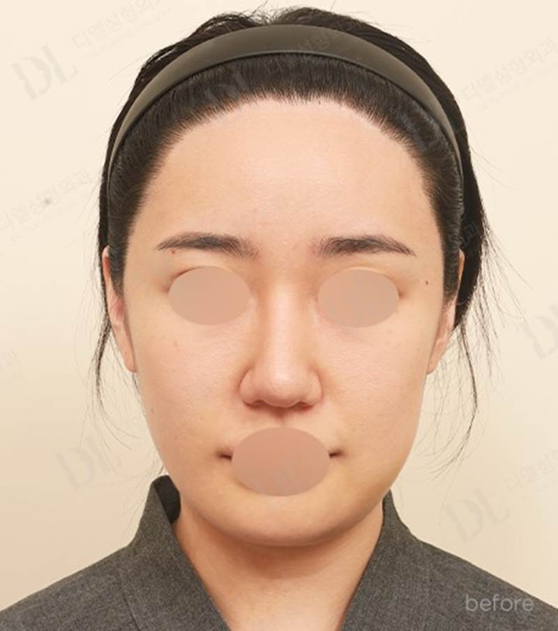
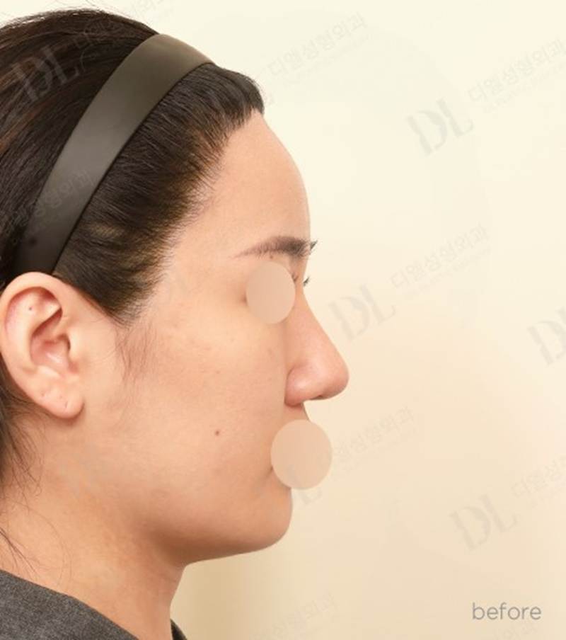
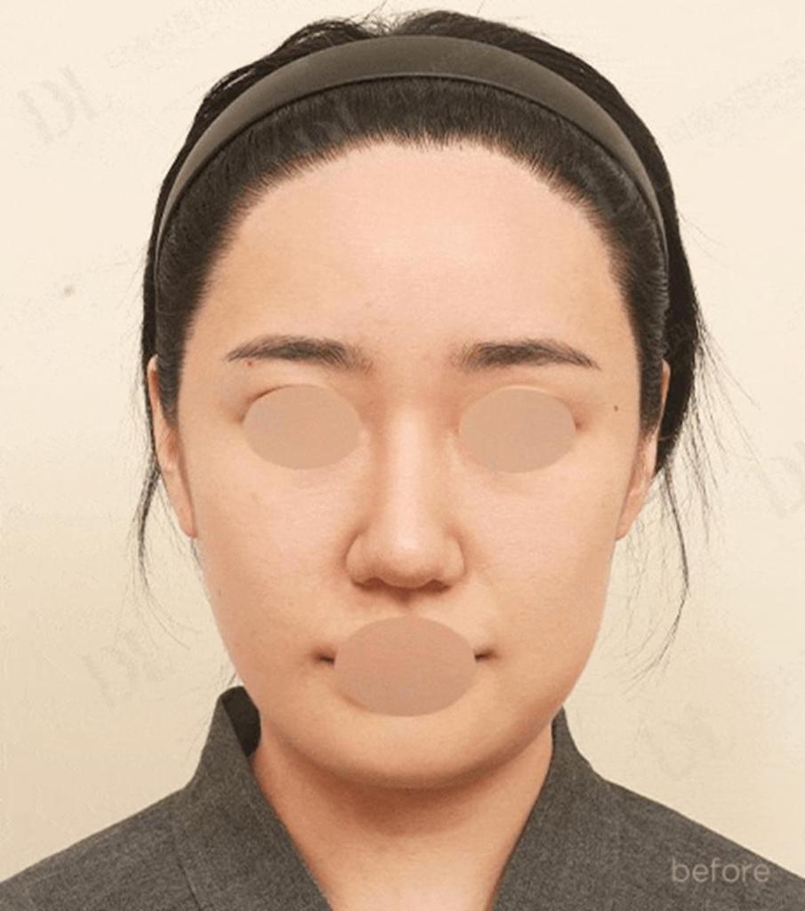
3-Area Facial Contouring + Paranasal Augmentation + Fat Grafting | Preoperative View
When the lateral cheekbones are prominent and the mandibular angles are wide, the face can appear broad from the front and more angular from the side. Zygoma reduction and mandibular angle reduction can reduce the skeletal width. However, reducing bone width alone may create several aesthetic imbalances if the existing soft-tissue volume is not considered.
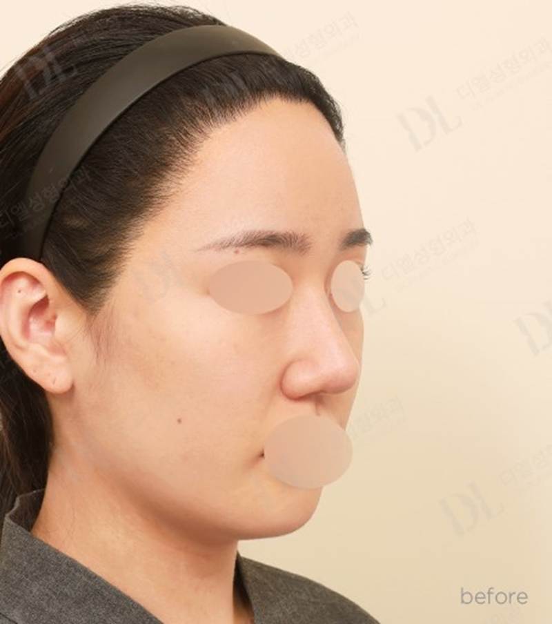
3-Area Facial Contouring + Paranasal Augmentation + Fat Grafting | Preoperative View
- • The midface may appear relatively flatter after the lateral cheekbone width is reduced.
- • The jawline may become slimmer, but the chin can still appear broad or visually flat if its width is not addressed.
- • A pre-existing nasolabial or paranasal depression may become more noticeable after the outer facial width is reduced.
For this reason, facial contouring should be designed by calculating both how much skeletal width should be reduced and where volume should be preserved or restored.
2. Precision Diagnosis and Surgical Planning
Before combining facial contouring surgery with paranasal augmentation, a detailed 3D CT analysis is performed to evaluate the facial skeleton. The main points reviewed on 3D CT include:
- • The degree of left and right zygomatic prominence and facial asymmetry
- • The angle and outward flare of the mandibular angles
- • The chin contour, projection, and relationship to the occlusion
- • The relationship between the facial skeleton and soft-tissue volume in the cheek and nasolabial regions
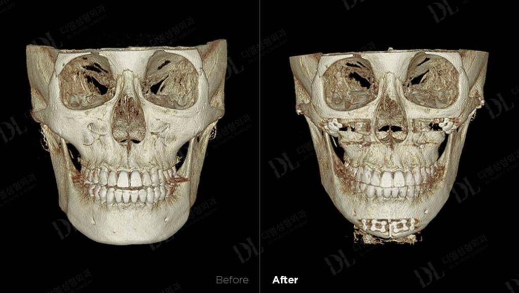
3-Area Facial Contouring + Paranasal Augmentation + Fat Grafting | Before → 2 Months After, 3D CT
A key part of diagnosis is determining whether facial width is primarily caused by the skeleton or by soft tissue such as fat and muscle. When skeletal reduction and volume restoration are planned together, the surgeon can estimate in advance where bone will be reduced and where soft-tissue volume may need to be preserved or restored, allowing a more precise prediction of the postoperative contour.
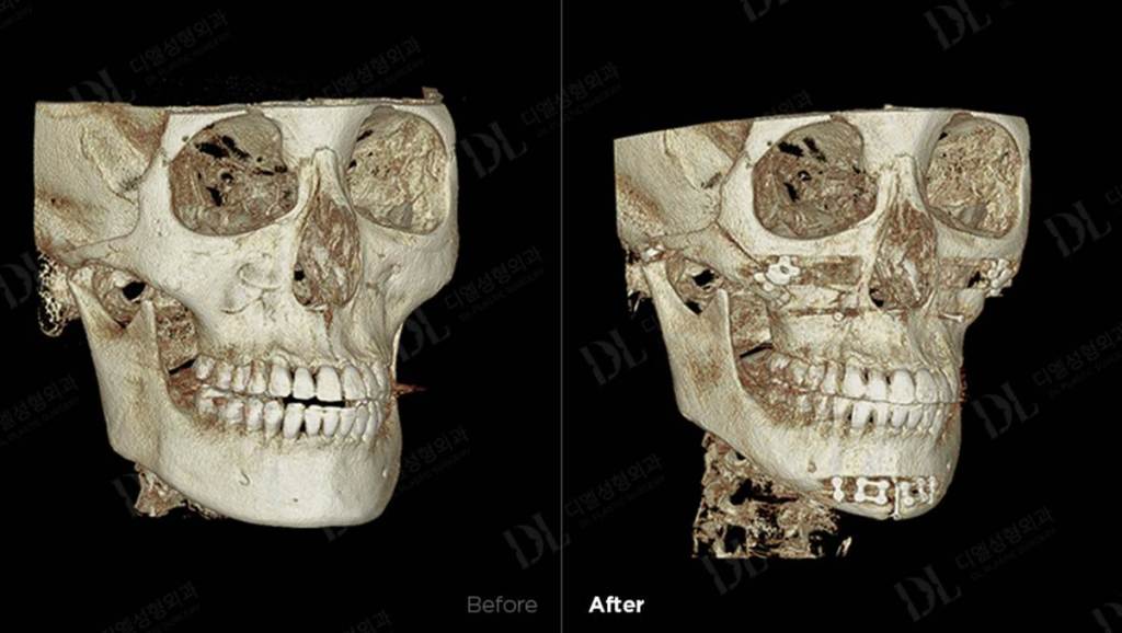
3-Area Facial Contouring + Paranasal Augmentation + Fat Grafting | Before → 2 Months After, 3D CT
On 3D CT, this case showed prominent lateral and 45-degree zygomatic projection. Zygoma reduction was planned by osteotomizing and repositioning the prominent zygomatic segment inward. Stable fixation with titanium plates and screws was used to maintain the planned position and reduce the risk of segment displacement or nonunion.
For the lower face, the prominent mandibular angle and broad jawline were addressed with long-curve mandibular angle reduction extending from the posterior angle toward the anterior jawline. The aim is to create a continuous contour without a secondary angle. The broad chin was then narrowed with a T-shaped genioplasty after carefully evaluating the nerve canal, allowing the lower facial width to be refined in proportion to the newly contoured jawline.
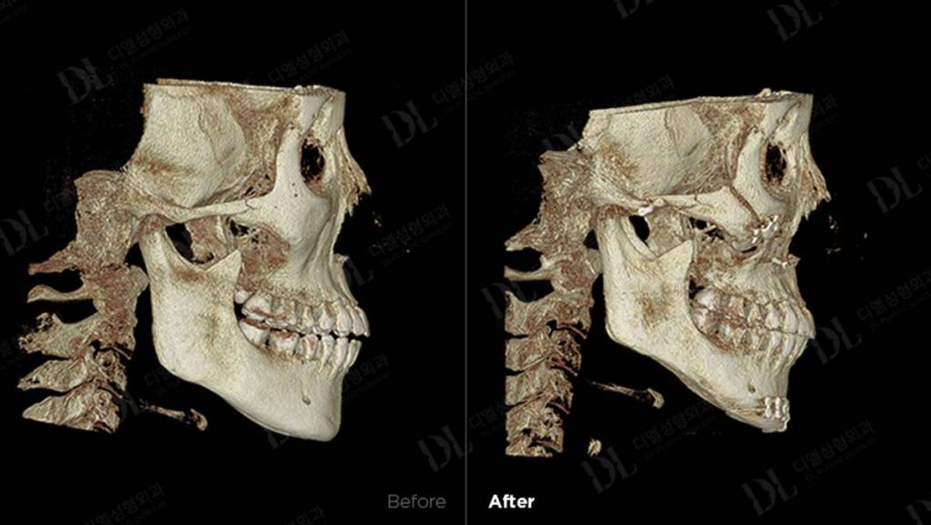
3-Area Facial Contouring + Paranasal Augmentation + Fat Grafting | Before → 2 Months After, 3D CT
For the recessed areas beside the nasal base and around the nasolabial region, a customized paranasal implant was used to improve central midface projection. Autologous fat grafting was also added to the relatively flat anterior cheek to restore subtle soft-tissue volume and improve the overall three-dimensional balance of the face.
3. A More Important Goal Than Simply Making the Face Smaller
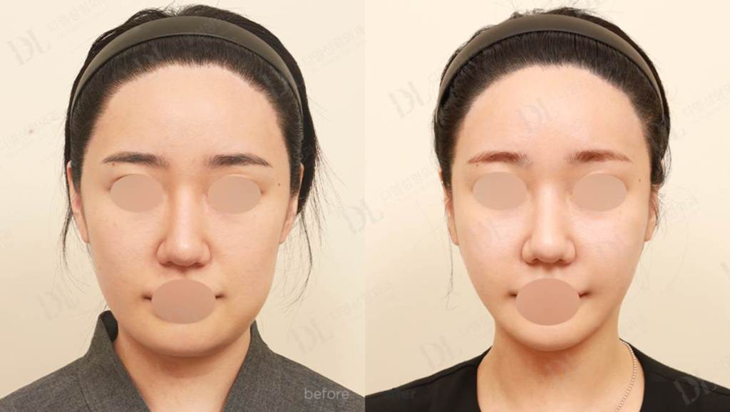
3-Area Facial Contouring + Paranasal Augmentation + Fat Grafting | Before → 2 Months After
At 2 months after surgery, the frontal view shows reduced width of the cheekbones and lower face, producing a softer overall facial outline. The previously prominent zygomatic and mandibular contours are less dominant, while the central facial region appears more defined.
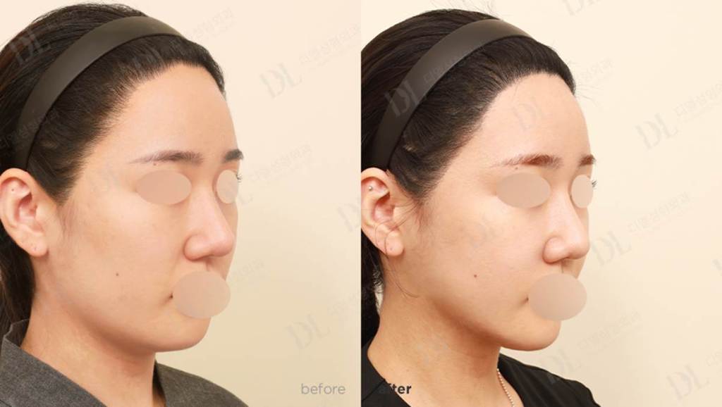
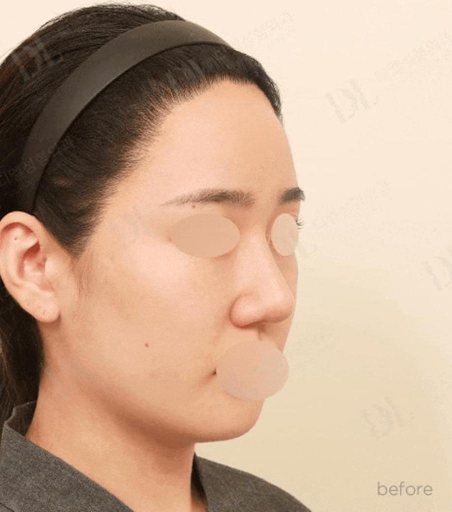
3-Area Facial Contouring + Paranasal Augmentation + Fat Grafting | Before → 2 Months After
From the lateral view, the preoperative chin position made the lower facial contour appear relatively blunt. After surgery, the chin shape was refined and the transition from the chin to the mandibular border became clearer, creating a more defined profile line.
4. Why Consider Facial Contouring Surgery and Paranasal Augmentation Together?
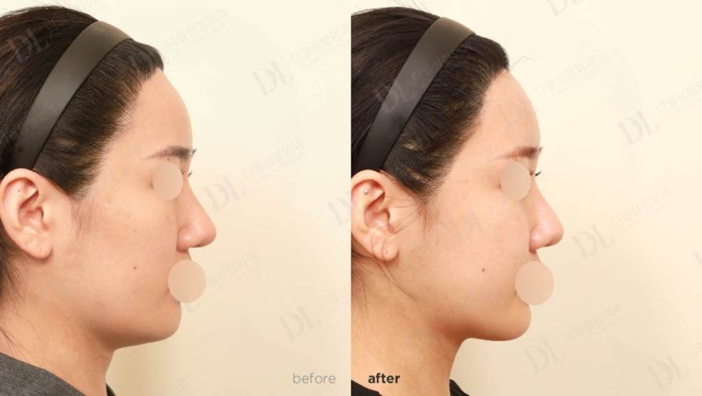
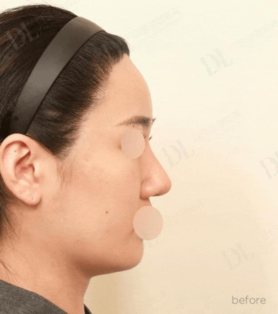
3-Area Facial Contouring + Paranasal Augmentation + Fat Grafting | Before → 2 Months After
Facial contouring surgery and paranasal augmentation may seem like opposite concepts. Facial contouring reduces excessive skeletal width and projection, while paranasal augmentation adds volume to the central midface beside the nasal base. In selected patients, however, these two approaches can complement each other.
When the outer facial framework is wide but the central midface is recessed, facial contouring alone may make the face appear relatively flatter. In this situation, reducing the outer contour while selectively supporting the center of the face can help preserve three-dimensional balance. Conversely, if the central midface already has sufficient skeletal and soft-tissue volume, unnecessary augmentation may make the nasal base area appear too prominent.
Therefore, paranasal augmentation should not be added simply because it is popular. It should be considered only after evaluating frontal, oblique, and lateral facial views together with CT findings to confirm that central volume support is actually needed.
This combined approach may be considered when:
- • Prominent cheekbones and mandibular angles make the outer face appear wide.
- • The overall facial outline appears broad or angular.
- • The chin appears short, broad, or relatively retruded.
- • There is concern that facial contouring alone may make the midface appear too flat.
- • The area beside the nose or around the paranasal region appears recessed.
- • Nasolabial depression and reduced midface volume are present together.
- • The goal is to reduce outer facial width while preserving natural three-dimensional facial volume.
5. Conclusion
The key principle of combining facial contouring surgery with paranasal augmentation is simple: reduce excessive angularity while preserving or restoring volume where it is needed. The objective is not merely to create a smaller face, but to achieve a balanced contour that remains three-dimensional from the frontal, oblique, and lateral views. For this reason, surgical planning should evaluate not only how much bone should be reduced, but also what volume should remain after the skeletal framework has been changed.
A natural facial shape is not created by maximum reduction. It is created when skeletal angles and facial volume remain in balance.
Frequently Asked Questions(FAQ)
Q. What is 3-area facial contouring surgery?
A. It generally refers to zygoma reduction, mandibular angle reduction, and chin surgery such as genioplasty. The exact combination depends on the patient’s anatomy.
Q. Why combine facial contouring with paranasal augmentation?
A. In selected patients, the outer facial framework is wide while the central midface is recessed. Reducing the outer width while selectively supporting the center can help maintain a three-dimensional facial balance.
Q. Can facial contouring make the face look hollow?
A. It can appear relatively flatter if pre-existing soft-tissue or midface volume deficiency is not considered during planning. This is why both skeletal structure and volume distribution should be evaluated before surgery.
Q. Why is 3D CT used before facial contouring surgery?
A. 3D CT helps evaluate cheekbone prominence, mandibular angle, chin anatomy, asymmetry, nerve location, and the relationship between the facial skeleton and the planned osteotomies.
Medical information notice: This article is for general educational purposes and does not replace an in-person consultation. Surgical indications, techniques, recovery, and risks vary according to individual anatomy and medical condition.
 DL Plastic Surgery