
Causes and Four Surgical Principles for Risk Reduction
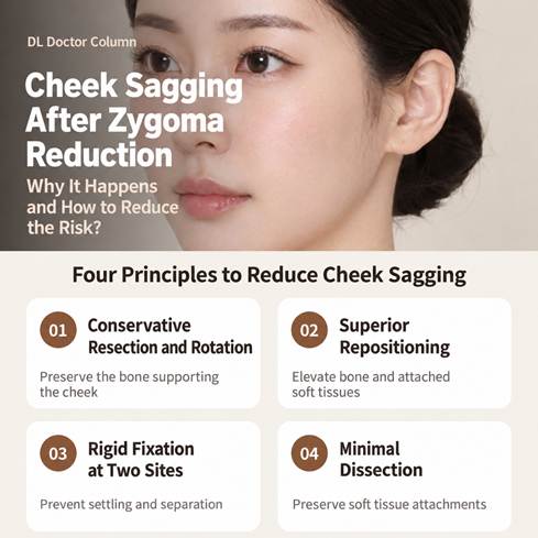
Figure 1. Overview of the four operative principles.
Abstract
Cheek ptosis is a frequently expressed concern in reduction malarplasty. The source article describes it as a potentially modifiable complication related to skeletal support, segment stability, and preservation of soft tissue attachments. This clinical review organizes the discussion around four operative principles: conservative bone resection with rotational reduction, superior repositioning, rigid fixation of the zygomatic body and arch, and limited dissection. Preoperative three dimensional computed tomography and assessment of skin elasticity, fat distribution, and previous procedures guide individualized planning. Accurate repositioning and stable union are central to maintaining cheek support; the extent of bone removal alone does not determine the aesthetic result.
Keywords reduction malarplasty; cheek ptosis; zygoma; L shaped osteotomy; rigid fixation; retaining ligaments
Key Answer
Cheek sagging after zygoma reduction is a possible complication, but it is not inevitable. The source article links the risk to loss of skeletal support, unstable fixation, and extensive soft tissue dissection. Its four prevention principles are conservative resection with rotation, superior repositioning, rigid fixation at the body and arch, and minimal dissection. Individual anatomy and tissue quality still influence outcomes.
What Is Zygoma Reduction Surgery
“Will my cheeks sag after zygoma reduction?” is one of the questions most frequently raised during consultation. Cheek descent is not presented in the source as an inevitable consequence of reduction malarplasty. Rather, its risk is influenced by the operative design, the method of fixation, and the handling of surrounding soft tissues.
The purpose of reduction malarplasty is to narrow excessive malar prominence while retaining a balanced three dimensional contour and adequate soft tissue support. This requires attention to the direction of skeletal movement as well as the amount of resection. The four principles discussed below provide a framework for this approach.
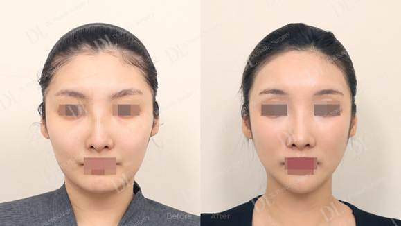
Figure 2. Clinical comparison reproduced from the first photograph in the source post.
The source describes minimal visible cheek descent without an additional lifting procedure.
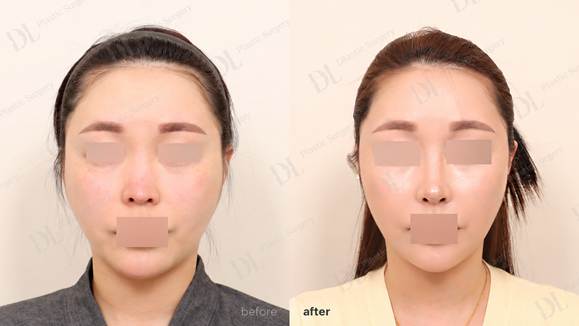
Figure 3. Clinical comparison reproduced from the second photograph in the source post.
Images are illustrative and do not establish comparative efficacy.
Why Cheeks May Sag After Zygoma Reduction
The relationship between the cheek and the zygoma can be compared with a curtain and its supporting rod. If the rod is excessively thinned, loosely mounted, or separated from the curtain, the curtain may descend. Similarly, excessive bone removal can diminish skeletal support; unstable fixation can permit inferior settling or nonunion; and extensive dissection can detach the soft tissue and muscle envelope from the underlying bone.
The source cites a three dimensional CT analysis of changes in the zygomatic summit after reduction malarplasty. In that analysis, the extent of bone resection primarily influenced medial displacement, whereas setback altered the summit position in superior, anterior, and medial directions [1]. These effects depend on the geometry of the mobilized segment and should not be reduced to a simple rule that greater resection produces a better result.
Excessive resection may weaken the supporting framework and make stable healing more difficult in the presence of muscular traction. Accordingly, the intended direction of movement and the quality of fixation deserve at least as much attention as the amount of bone removed.
Principle 1 Conservative Resection and Rotational Reduction
Bone removal alone does not necessarily produce a substantial reduction in facial width. The source describes medial rotation of the lateral zygomatic segment around the anterior osteotomy, analogous to closing a door around its hinge. Appropriate rotation can narrow a laterally flared arch while limiting the amount of supporting bone that must be removed.
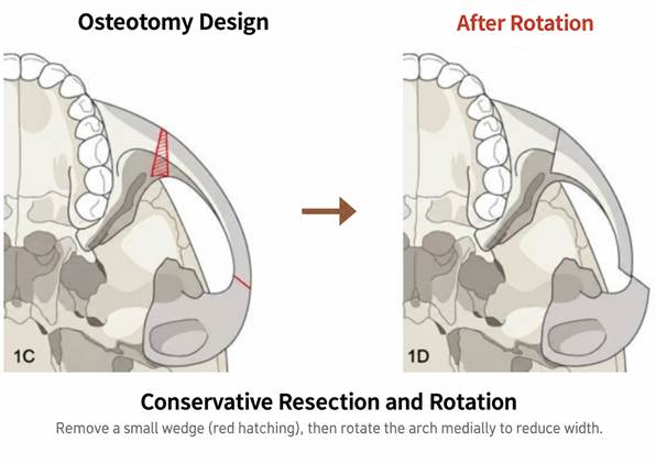
Figure 4. Limited wedge resection followed by medial rotation of the zygomatic arch.
The osteotomy configuration influences how this rotation can be achieved. The source favors an L shaped osteotomy because it provides a longer bone contact surface and accommodates stable fixation and multidirectional repositioning. Its comparison with an I shaped osteotomy is summarized in Table 1. These characteristics are presented as the rationale for the technique rather than as numerical estimates of comparative complication rates.
Table 1 Osteotomy characteristics described in the source article
| Characteristic | I shaped osteotomy | L shaped osteotomy |
| Cut configuration | Single linear cut | Horizontal and vertical components |
| Bone contact | Relatively narrow; potential for displacement or nonunion | Longer and broader contact surface |
| Fixation | More limited in the source comparison | Rigid fixation at the body and arch |
| Movement | Primarily medial translation | Medial and superior movement with rotation |
| Resection requirement | May require greater resection for a comparable reduction | Rotation can reduce the required resection |
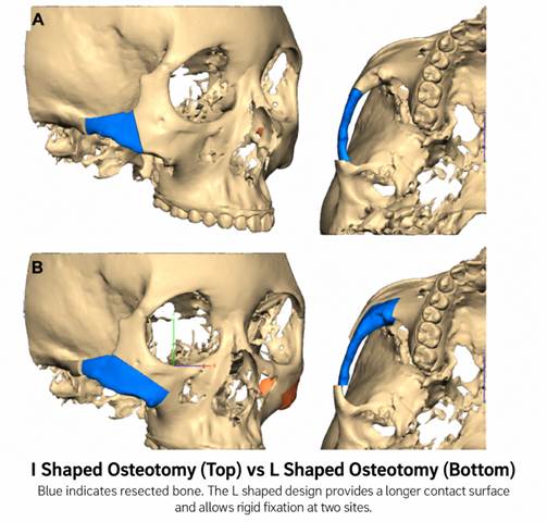
Figure 5. I shaped and L shaped osteotomy configurations. Blue areas indicate resected bone.
The width of an L shaped resection may be varied to create a wedge according to the pattern of malar prominence. This geometry permits controlled rotation and can increase contact between the repositioned bone surfaces. Preserving a supportive bony framework and obtaining stable union are intended to reduce the tendency toward postoperative cheek descent.
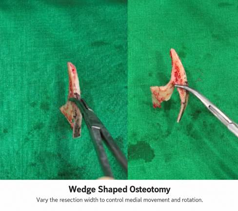
Figure 6. Wedge shaped bone resection used to control rotation and medial displacement.
Even within an L shaped design, the location and orientation of the cut affect the rotational result. The source uses the zygomaticofacial foramen, through which a sensory nerve to the cheek emerges, as an anatomical reference. In the illustrated CT plan, the blue lines represent a less favorable rotational direction or insufficient rotation, while the red lines represent the intended design. Preoperative three dimensional CT is used to map the nerve location and individual bony anatomy before selecting the osteotomy.
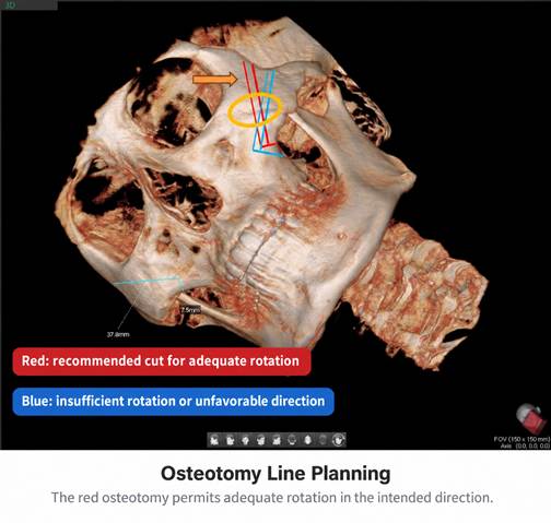
Figure 7. CT based osteotomy planning relative to the zygomaticofacial foramen.
The red and blue cuts illustrate different rotational effects.
Principle 2 Superior Repositioning
After an L shaped osteotomy, the mobilized zygoma is repositioned medially and superiorly. Provided that the relevant soft tissue attachments are preserved, the overlying cheek can move with the elevated skeletal segment. This is analogous to remounting a curtain rod at a higher level.
The malar summit, designated MMP in the source figures, is moved superomedially rather than simply upward. In patients with prominent zygomas, this point may lie inferolateral to the desired position. Superomedial movement can narrow the face while maintaining anterior cheek fullness and facial depth.
Posterior repositioning of the prominent segment may be incorporated to modify the malar highlight. The resulting movement of the summit reflects the combined segment geometry, rotation, and translation. The intended outcome is a narrower contour with a supported cheek envelope and a more favorable highlight position.
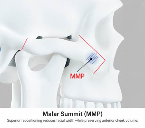
Figure 8. The malar summit designated MMP in the source image.
Principle 3 Rigid Fixation at the Body and Arch
An appropriately designed osteotomy cannot compensate for inadequate fixation. Instability may permit segment descent or nonunion and can be associated with cheek ptosis and pain. The source author describes these findings among the problems encountered during revision consultations.
The technique described uses titanium plates to secure both the anterior zygomatic body and the posterior arch. The masseter attaches to the zygomatic complex and applies substantial force during mastication. Insufficient fixation, or fixation at only one site in a construct that does not adequately resist these forces, can allow the repositioned segment to settle inferiorly.
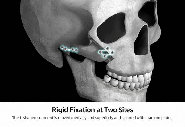
Figure 9. Fixation at the zygomatic body and arch after medial and superior repositioning.
Three technical considerations are emphasized. A saw with a defined resection width helps reproduce the planned bone removal.
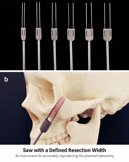
Figure 10. Saws with defined resection widths and an example of their application.
Prebent plates are used to maintain the planned position while avoiding additional intraoperative bending; the source notes that bending can affect metal strength.
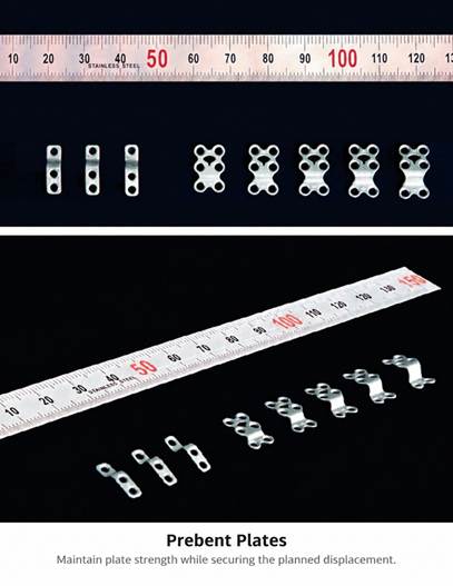
Figure 11. Prebent plates for fixation at the planned segment position.
Screw placement is selected in areas with sufficient bone thickness because the zygoma is not uniformly thick.
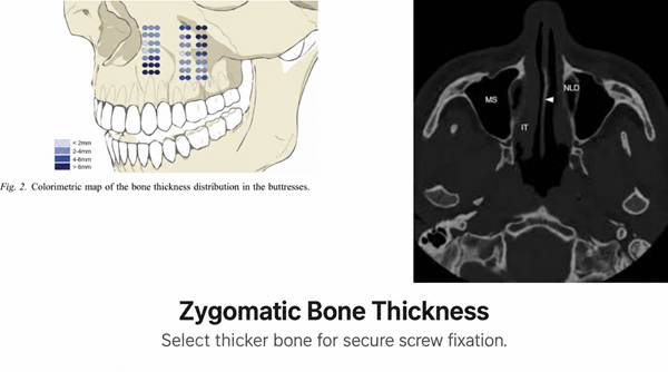
Figure 12. Variation in zygomatic bone thickness and selection of screw sites.
The posterior arch is described as broad anteriorly, narrow centrally, and wider again posteriorly. Moving the osteotomy posteriorly can improve lateral narrowing, but the temporomandibular joint limits how far posteriorly the cut can safely be placed. The source therefore locates the posterior osteotomy immediately anterior to the joint.
A beveled osteotomy increases the contact surface between bone ends. The source describes this as a means of promoting union while reducing palpable step deformity and anterior malar hollowing [5]. Stable primary fixation is a central component of the strategy to reduce postoperative descent and the need for revision.
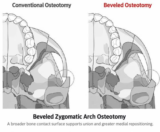
Figure 13. Conventional and beveled posterior arch osteotomies.
Principle 4 Minimal Dissection
Cheek soft tissue does not simply rest on the underlying bone. Retaining ligaments and muscular attachments help maintain its position. The zygomatic retaining ligament extends from the zygomatic region to the cheek skin, while masseteric retaining ligaments are associated with the anterior border of the masseter. The zygomaticus muscles and masseter also have attachments to the zygomatic complex.
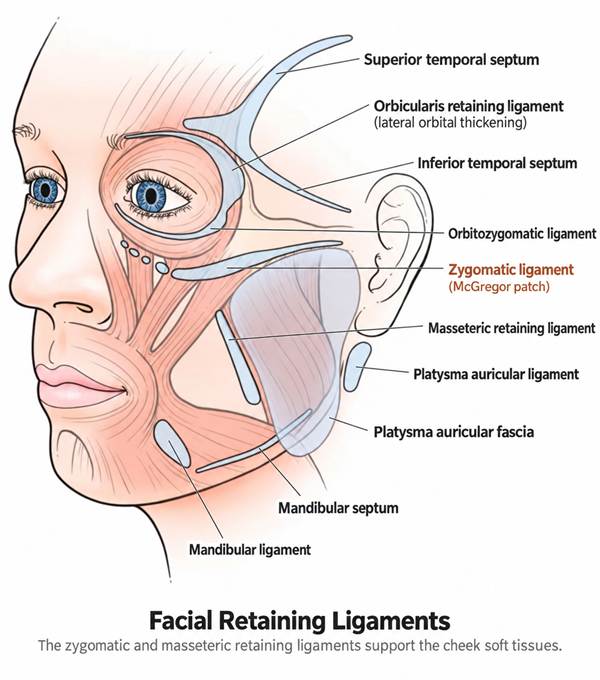
Figure 14. Facial retaining ligaments and related septa supporting the soft tissue envelope.
Extensive release of these structures may allow the cheek envelope to descend, much as a curtain falls when detached from its supporting rod. The source therefore advocates limiting dissection to the exposure needed for skeletal manipulation and preserving muscle attachments whenever possible.
The functional aim is to maintain the connection between the skeletal segment and the soft tissue envelope so that superior repositioning of the bone can also support the cheek.
Soft Tissue Assessment and Adjunctive Procedures
Patients undergoing similar skeletal procedures may have different soft tissue responses. Skin thickness, the amount and distribution of facial fat, and tissue elasticity influence how closely the external contour follows skeletal movement. A history of fillers or fat grafting should also be included in the assessment.
Patients in their forties and fifties may have reduced skin elasticity and a greater tendency toward postoperative cheek descent. Reduction malarplasty may still be considered, but lifting procedures or a facelift may be incorporated when clinically indicated.
The source discusses buccal fat removal and facial liposuction when excess cheek or jowl fat contributes to a broad or heavy appearance.
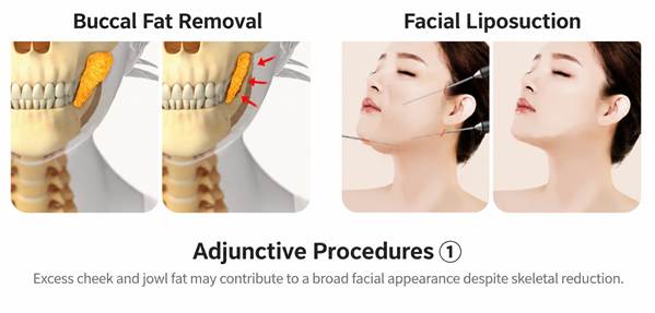
Figure 15. Adjunctive buccal fat removal and facial liposuction.
The source also describes fat grafting to the anterior malar region and temples to improve volume and contour. Paranasal augmentation may be considered when there is hollowing near the nasolabial base, because access can overlap with the malar procedure. These adjuncts are individualized options, rather than a requirement for every patient.
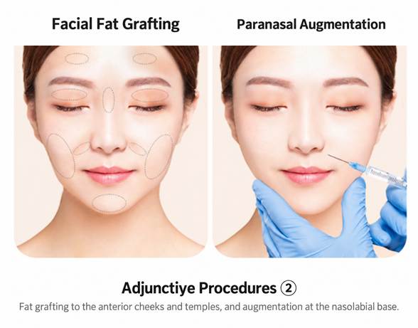
Figure 16. Adjunctive facial fat grafting and paranasal augmentation.
Does Zygoma Reduction Always Cause Cheek Sagging
No. Cheek sagging is a potential complication rather than an inevitable outcome. Surgical planning, stable bone support, preservation of soft tissue attachments, and the patient’s tissue quality are relevant to the risk. The source does not provide a numerical complication rate.
Does Removing More Bone Give a Better Result
Not necessarily. Excessive resection can reduce skeletal support. The source emphasizes controlled rotation and repositioning with limited bone removal to achieve a narrower contour while maintaining support.
Can Zygoma Reduction Be Performed After Age Forty
Age alone does not determine suitability. The source recommends assessing skin elasticity, facial fat, prior procedures, and the possible need for an additional lifting procedure. A clinical evaluation is required to select an appropriate approach.
Is a Facelift Necessary With Zygoma Reduction
A facelift is not described as a routine requirement. The source presents lifting as an individualized option when tissue laxity warrants it. The clinical photographs do not establish that every patient can avoid a lifting procedure.
What Should Patients Ask During Consultation
Consultation should address four practical questions: Is the direction of rotation and superior or posterior movement planned as carefully as the amount of resection? How will the body and arch be fixed, and at how many sites? What dissection is necessary, and how will soft tissue attachments be preserved? Have skin elasticity, facial fat, and previous procedures been evaluated?
The source directs readers to the third installment of its series for recovery time, swelling, diet, and facial washing guidance.
Discussion
The operative concepts described here combine preservation of skeletal support, controlled three dimensional repositioning, stable fixation, and protection of the soft tissue envelope. Their shared rationale is that a narrower facial skeleton must continue to provide an appropriate platform for the overlying cheek.
The clinical photographs reproduced from the source are illustrative. The source describes minimal visible cheek descent after malar reduction without an additional lifting procedure. These images do not establish a complication rate, a controlled comparison, or the magnitude of risk reduction. Individual anatomy and tissue quality remain relevant to interpretation of the result.
Conclusion
The source article identifies four principles for reducing the risk of cheek ptosis after reduction malarplasty: conservative resection with medial rotation, superior repositioning, rigid fixation at the zygomatic body and arch, and minimal dissection. The central emphasis is accurate movement and stable support, with individualized soft tissue assessment, rather than maximal bone removal.

