
Individualized design for natural nostril contours and less visible scarring
Consultations about the nasal base often begin with familiar concerns: “My tip has been raised, but my nostrils still look flared.” “My nose becomes wider when I smile.” “I have heard that nostril reduction leaves scars.

Medical team photograph from the source column.
The alae, or outer nostril wings, help define the width of the lower third of the nose. Even with a refined bridge and tip, pronounced flare can make the nose appear broad or bulky. Yet concerns about scarring often make patients hesitate to consider alar base reduction.
This clinical column explains why the nasal base may look wide, how surgical design changes with anatomy, the principles used to minimize visible scars, and the role of an alar lift when the two sides sit at different heights.
Anatomical considerations in Korean and other Asian patients
Many conventional descriptions of alar reduction were developed around Western nasal anatomy. Some Korean and other Asian patients present with a different combination of features: a broad nasal base, outward flare, thicker skin and subcutaneous tissue, relatively broad or less projected tip cartilage, and ample vestibular skin inside the nostrils.
These features can influence the design. Limited tip projection may contribute to the impression of outward flare, while the amount of internal skin affects how the base can be narrowed. However, anatomy varies substantially within every population. Ethnicity is a starting point for discussion, not a substitute for individual examination.
The source column notes that published reviews describe differences in the frequency of broad alar attachments and outward flare across populations. The practical implication is to match the operation to the individual nose rather than apply a single textbook pattern to everyone.
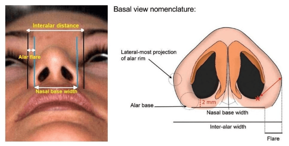
Figure 1. Basal-view landmarks distinguishing interalar distance, flare, and nasal base width.
Two different reasons the nasal base can appear wide
Alar flare
Alar flare is the rounded outward projection of the nostril wing beyond its attachment to the face. The source describes extension of more than approximately 2 mm beyond the attachment as a reference for assessing flare. This is an assessment aid rather than a universal indication for surgery.
A broad alar base
A broad base means that the attachments themselves sit relatively far apart. The distance between the inner eye corners is a traditional proportional reference. The source mentions approximately 31–33 mm, but neither that number nor a fixed ratio should be treated as an ideal for every face.
Nostril shape also matters. On the basal view, an obliquely oriented, pear-shaped opening may be an aesthetic preference, whereas a horizontally oriented opening can contribute to a broad, flat appearance. Natural variation and the patient’s preferences should guide the discussion.
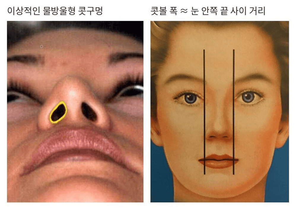
Figure 2. Nostril shape and facial proportions.
Korean labels refer to a pear-shaped nostril and the comparison between alar width and inner-eye distance.
Flare is addressed principally near the external alar crease. Base width may require reduction at the nostril floor, also called the sill. When both are present, external and internal components may be combined. The incision location therefore depends on the anatomical problem: sill reduction alone may leave outward flare, while excessive external excision can distort the contour without adequately correcting base width.
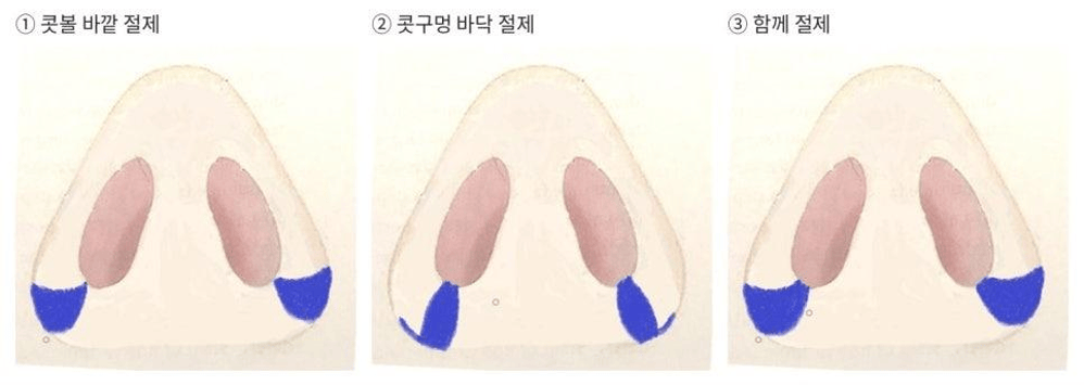
Figure 3. Excision regions. Left to right: external alar excision, nostril sill excision, and combined excision.
Why alar reduction is often the final step of rhinoplasty
In the approach described in the source, the design is planned before surgery, but the final extent is decided after bridge and tip work and closure of the main incision. Changes in tip projection can change apparent flare. In some patients with limited projection, tip surgery alone may reduce the amount of alar reduction required.
Measurements and markings are made separately on each side before local anesthetic injection, because injected fluid can swell the tissue and alter its appearance. The sides are rarely identical, so removing the same amount from each side is not necessarily the best route to improved balance.
How design changes with the pattern of flare
For predominantly flared alae, a crescent-shaped excision along the outer crease can reduce outward curvature. The source describes three patterns based on the position of greatest convexity when viewed from below.
Type 1 flare near the lower attachment
When the most prominent point is low and close to the alar attachment, the flared segment is relatively limited. A short, conservative excision helps preserve the natural curve. Excessive removal can flatten or distort it.
Type 2 flare near the level of the attachment
When the maximum convexity lies near the level of the attachment, the design may extend farther upward along the crease. The exact extent follows the individual contour.
Type 3 flare extending toward the tip
When the most convex area lies higher toward the tip, a larger area may contribute to flare. The source associates this pattern with some broad noses, lower tip projection, and larger nostrils. It may require a more extensive design, while still preserving the remaining tissue and contour.
The patterns can differ between the two sides. One side may resemble Type 2 and the other Type 3, requiring separate markings. The key principle is the shape that remains. Eliminating every trace of flare can leave an unnaturally straight alar wall. Retaining a gentle curve may also help conceal the scar within the crease.
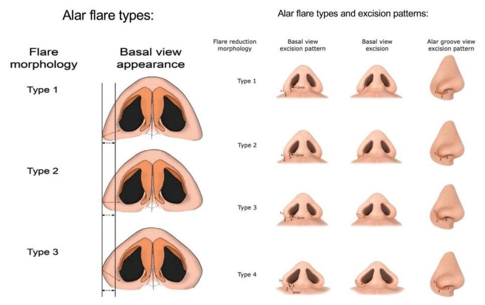
Figure 4. Alar flare morphology and corresponding excision patterns.
The source figure includes a fourth illustrated pattern in addition to the three patterns discussed in the text.
A broad base with little outward flare
When the attachments are wide but the wings are not markedly flared, a wedge-shaped reduction at the nostril sill may help move the attachments inward. This can narrow the lower nose and alter a round opening toward a more oval outline.
Sill reduction alone does not necessarily correct external flare. It also reduces the dimensions of the opening, so an already small nostril calls for particular caution to preserve breathing.
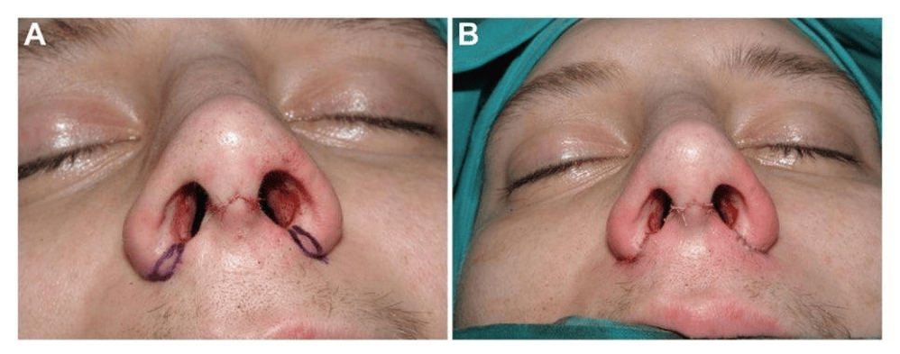
Figure 5. Clinical operative photographs accompanying the source discussion of nasal base reduction.
Combined flare and base width
Outward flare and a broad base often occur together. In the source approach, an external alar excision is connected with a sill excision to address both components. Published clinical experience discussed in the source describes improvements in flare and nostril orientation, but individual results vary. Scarring, residual asymmetry, and contour changes remain possible.
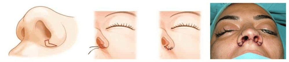
Figure 6. Combined alar and nostril sill reduction illustrated in the source column.
Conservative external excision with internal reduction and cinching
A markedly broad, flared base may require excessive external removal if treated from the outside alone. Potential consequences include visible scars, notching of the alar margin, and loss of the natural alar-cheek groove.
Suture-only narrowing also has limits. In a strongly flared nose, pulling the bases together can bunch the sill or exaggerate the rounded alar contour. A symmetric cinching force may not independently correct unequal tissue shape on each side.
The source describes the combined method reported by Oh and colleagues in 2010. Its components include limited external excision to address flare, relatively greater internal vestibular skin reduction, limited mobilization of the attachments, and a cinching suture to help bring the bases inward.
In that reported method, the average external excision width was less than 3 mm, and internal excision was approximately two to three times greater. These are descriptions of a particular technique, not dimensions that can be applied to every patient. The cinching suture passes between the alar bases through tissue near the upper lip and the base of the columella. Its placement and tension influence nostril shape.
The components serve different purposes: the external excision addresses flare, while internal reduction and cinching address width. Removing appropriate internal tissue may reduce bunching and the tension needed to bring the bases inward. More force is not inherently better; excessive tension may cause temporary upper-lip swelling or discomfort.
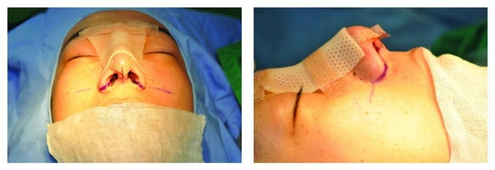
Figure 7. Operative photographs accompanying the discussion of internal reduction and cinching
Other anatomical situations
If the alar tissue itself is thick, selected internal excision may reduce bulk without an external skin incision. If the base is broad but flare is modest, a limited-incision suture technique may be considered. Neither approach is suitable for every nose, and a smaller external incision does not eliminate all surgical risks.
Uneven alar height and the role of an alar lift
Sometimes the main difference is vertical position rather than width. One attachment sits lower, making the nasal base appear tilted. Possible contributors include unequal maxillary height, scar traction after surgery or injury, and asymmetry of tip cartilage or alar soft tissue.
A tilted base can make the nose appear uneven even after other deviation has been addressed. Assessment therefore includes the surrounding face. Frontal photographs can help compare a line through the pupils with a line connecting the lower alar bases.
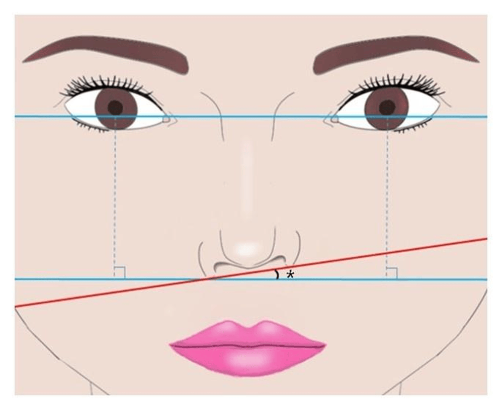
Figure 8. Assessment of alar-base tilt relative to the pupil reference line.
The source describes an alar lift performed on the lower side near the end of rhinoplasty. A pointed ellipse of vestibular skin immediately inside the nostril sill, with a minimal amount of underlying tissue, is removed and closed without extensive deep dissection. The source reports an average design of approximately 7 by 5 mm; the actual design must be individualized.
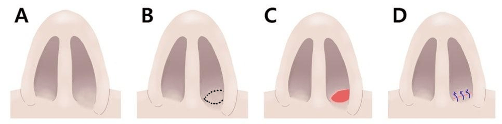
Figure 9. Schematic sequence of the internal alar lift technique.
The internal location avoids an external skin incision for this component. It does not mean that no scar forms or that infection, contraction, or other complications are impossible. The source reports improved height differences and no procedure-related complications in the cited series, which should not be interpreted as a guarantee for all patients.
An alar lift primarily addresses height. A separate difference in width may require a different reduction design. Severe scar-related retraction may make the technique unsuitable, and internal reduction must avoid excessive narrowing of the nostril.
Six principles for minimizing visible scars and distortion
Keep external removal conservative
External tissue should be removed only to the extent needed for the flare being treated. Where appropriate, internal reduction and cinching can contribute to width correction. Scar length and wound tension are important considerations, alongside skin characteristics and healing biology.
Respect natural creases
The incision may follow the junction between the ala and cheek and continue into the groove between the sill and upper lip. Placement is adjusted to anatomy and skin type. The source discusses differing preferences for an incision within the crease or slightly above it. Neither position is universally appropriate. Excessive superior extension may make the scar more conspicuous and put local tissues at risk.
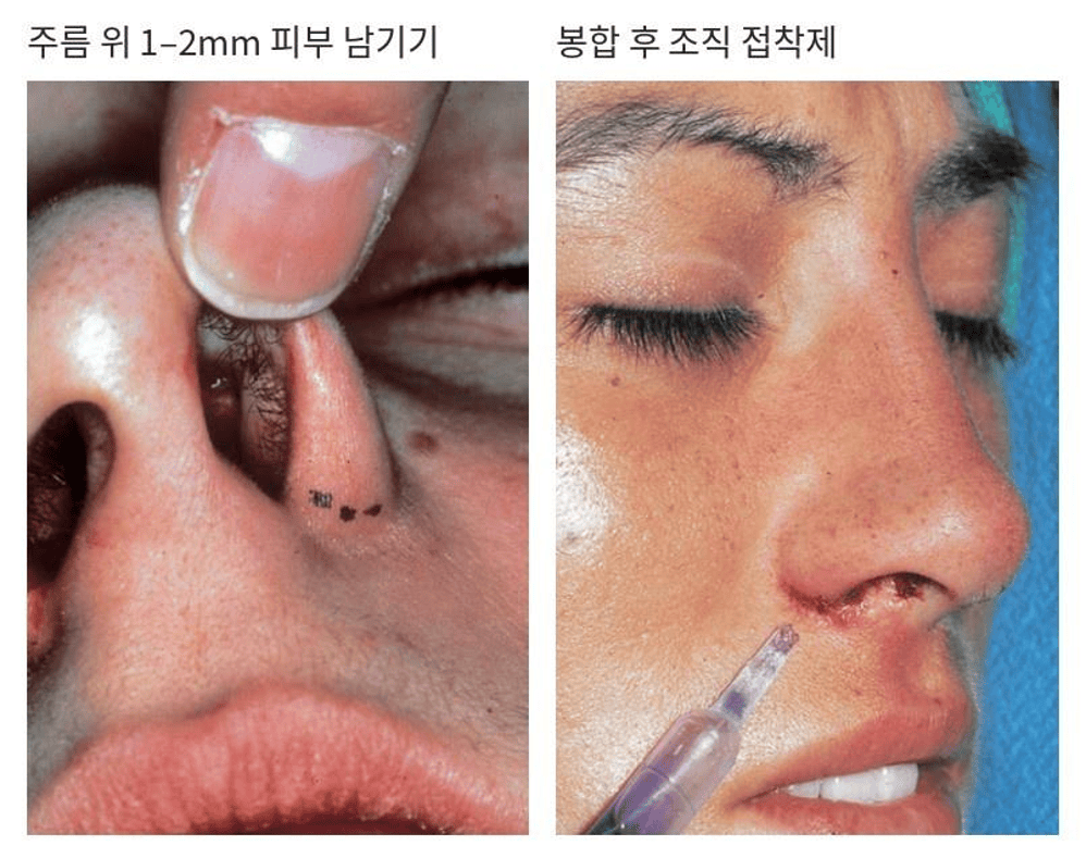
Figure 10. Incision and closure details.
Korean labels describe retaining 1–2 mm of skin above the crease and precise alignment of the wound edges.
Preserve the nostril sill contour
The source describes retaining approximately 2 mm of a vertical sill segment in the relevant design to help prevent notching. This is a technique-specific detail. When only flare is being treated, unnecessary extension into the nostril can distort the opening.
Address the more flared side conservatively
The described approach treats the more flared side first, then adjusts the opposite side to match. Conservative removal helps avoid overcorrecting the less flared side. Reconstructing an over-resected ala can be difficult; a small residual concern may be reassessed after healing before further treatment is considered.
Align the wound edges accurately
Careful approximation, with appropriate edge alignment and eversion, helps avoid a step that can cast a shadow and highlight a scar. Excessively tight sutures can leave marks. The source describes fine sutures and removal at around five to seven days, but the treating surgeon should determine timing for the individual wound.
Protect the healing area
The source recommends avoiding rubbing or manipulating the alar base for approximately two weeks. Follow the surgeon’s specific cleansing and ointment instructions, attend follow-up, and begin scar management when advised. Healing varies, and scar visibility cannot be predicted from technique alone.
Setting realistic expectations
Alar base reduction involves changes measured in millimeters. The reduction in overall width may be smaller than the amount of tissue removed. Much of the visible improvement can come from changes in curvature and nostril orientation rather than a large numerical reduction.
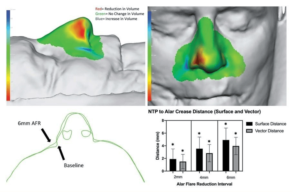
Figure 11. Source research illustration showing surface changes and measurements associated with alar flare reduction.
Narrowing can also reduce the nostril opening. Patients with small openings require a particularly conservative plan that considers airflow. The intended width should suit the tip, bridge, facial proportions, and the patient’s preferences. A somewhat wider base may be harmonious on a wider face.
If the need for reduction is uncertain, staging can be considered. The source suggests reassessment approximately six to twelve months after rhinoplasty, once swelling has sufficiently settled. A later procedure may be possible under local anesthesia, depending on the patient and the extent of correction.
Questions to bring to the consultation
- Is my concern outward flare, a broad attachment, or both?
- Have nostril size, orientation, and shape been assessed?
- Are the two sides different in width, height, or both?
- If tip surgery is planned, when will the final extent of reduction be determined?
- How will external excision, internal reduction, and any cinching suture contribute to the plan?
- How do facial asymmetry or planned facial contouring procedures affect the assessment?
The aim is a nasal base that suits the individual face while preserving its natural contour and function. Conservative design and careful closure seek to minimize visible scars, but no operation can promise scar-free healing or perfect symmetry.

