
Three types of cheekbone prominence and individualized surgical planning
Cheekbone reduction surgery, also called zygoma reduction or reduction malarplasty, changes the position and, when appropriate, the shape of prominent cheekbones. Planning depends on where the projection occurs: along the lateral arch, around the area seen at a 45-degree angle, or across the cheekbone complex.
A wide-looking face does not automatically mean the cheekbone is unusually thick. Outward flaring, bone position, jaw width, and soft tissue all influence the appearance. The appropriate approach requires an individual assessment.
Which Cheekbone Type Do You Have A Quick Answer

Clinical introduction. Photograph from the original article.
Lateral prominence mainly increases width near the ears. 45-degree prominence is most noticeable from an oblique view. Overall prominence combines a broad cheekbone outline with a relatively flat midface. These are descriptive patterns used in this article; photographs alone cannot establish a surgical diagnosis.
- Width near the ears: the lateral zygomatic arch may be the main concern.
- A projecting contour in three-quarter views: the zygomatic body may need assessment as well as the arch.
- A broad, relatively flat midface: narrowing must be balanced against preserving forward cheek projection.
Which Cheekbone Type Do You Have A Quick Answer
Lateral prominence mainly increases width near the ears. 45-degree prominence is most noticeable from an oblique view. Overall prominence combines a broad cheekbone outline with a relatively flat midface. These are descriptive patterns used in this article; photographs alone cannot establish a surgical diagnosis.
- Anterior cheek volume
The fullness below the eyes contributes to facial dimension. This visible volume also depends on soft tissue, not only bone. Preserving it is an important consideration when planning reduction. - The 45-degree cheekbone area
This is the contour most noticeable in a three-quarter view. “45-degree cheekbone” is a descriptive aesthetic term, not a separate bone. - The lateral cheekbone or zygomatic arch
The arch extends toward the area in front of the ear and contributes to the outer width of the midface. - These areas should be assessed together. Narrowing the lateral outline while removing too much anterior projection may produce an unbalanced result.

Three areas of the cheekbone.
1 Anterior cheek volume. 2 The 45-degree cheekbone area. 3 Lateral cheekbone or posterior arch.
The source describes reducing areas 2 and 3 while preserving or restoring anterior fullness.
What Are the Three Types of Cheekbone Prominence
The following framework summarizes the three patterns described in the original clinical article. Some patients have a mixture of patterns, and the labels do not prescribe a standard amount of bone removal.
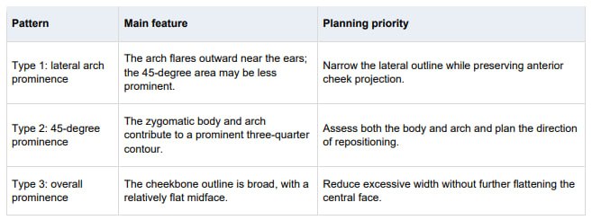
Type 1 Lateral Arch Prominence
In this pattern, the outer arch is the main contributor to width. The area closer to the front of the cheek may be less prominent, creating a visible transition between the anterior cheek and the wider outer outline.
The source article describes a rotation-focused approach with limited resection of the zygomatic body. The amount of movement and the fixation plan still depend on the individual anatomy.

Type 1 lateral arch prominence. Image credit as listed in the original article:
NMIXX, Instagram. An illustrative example, not a surgical diagnosis or treatment result.
In this pattern, the prominence involves the zygomatic body as well as the arch. It is often easiest to see in three-quarter photographs rather than in a straight-on view alone.
Addressing only the outer arch may not resolve projection from the zygomatic body. The source article describes combining resection with inward and posterior repositioning, tailored to the existing cheek volume and shape.

Type 2 45 degree prominence. Image credit as listed in the original article: Kim Shin-rok, Instagram.
An illustrative example, not a surgical diagnosis or treatment result.
Type 3 Overall Prominence with a Broad, Flat Midface
This pattern combines a broad cheekbone outline with less forward facial projection. A wide jaw may also contribute to the overall outline.
The source article emphasizes inward rotation while limiting posterior movement to avoid making an already flat midface look flatter. This is a planning principle within the source framework, rather than a universal recommendation for every broad face.

Type 3 overall prominence. Image credit as listed in the original article: Park Kyung-lim, Instagram. An illustrative example, not a surgical diagnosis or treatment result. Visible broadcast text: Monday, October 5, at 10 PM; the remaining text is cropped in the original.
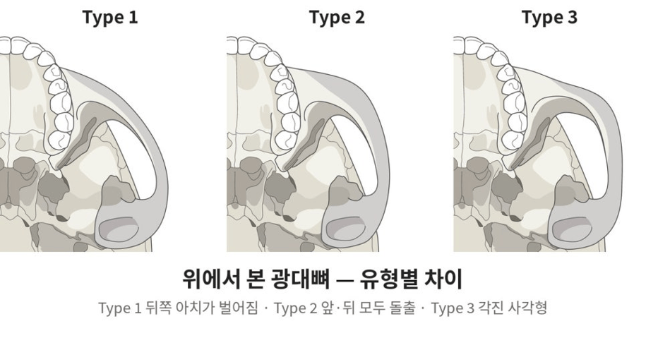
Top down comparison of cheekbone types. Type 1: outward-flaring posterior arch.
Type 2: anterior and posterior prominence. Type 3: a more angular, square configuration.
How Does Cheekbone Reduction Surgery Differ by Type
The key variables are where to make the bone cuts, whether to remove bone, and how to rotate and reposition the segments. The procedure name alone does not describe an individual surgical plan.
The original article describes an L-shaped osteotomy through an intraoral incision, combined with an arch osteotomy accessed through a small incision in the sideburn region. An osteotomy is a controlled surgical bone cut that permits repositioning. Other techniques also exist.
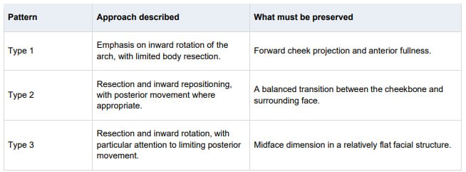
Published techniques also distinguish between prominence limited to the arch and prominence involving the cheekbone body. For example, one study evaluated a selected group with marked arch prominence and normal body prominence. Its findings do not establish a single best technique for all patients. Read the study abstract on PubMed.
Bone fixation and healing are part of the plan, not afterthoughts. Patients should ask how the repositioned segments will be stabilized and how healing will be assessed.
Why Is 3D CT Used Before Cheekbone Reduction
3D CT helps the surgeon assess the position, shape, and symmetry of the bones and plan osteotomy locations. It provides information that surface photographs cannot show, although it cannot by itself predict the final soft-tissue appearance.
A published technique describes CT-based planning of zygomatic arch osteotomies, highlighting the importance of matching the planned cut locations on both sides. This supports the role of imaging in planning; it does not mean imaging eliminates surgical risk. Read the CT planning study abstract.
The consultation should also address skin thickness and laxity, fat distribution, muscle volume, asymmetry, and previous filler, fat grafting, or facial surgery. The surgeon determines what imaging is appropriate for the patient.
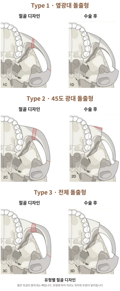
Osteotomy design by cheekbone type.
Each row shows the osteotomy design on the left and the postoperative position on the right.
Top: Type 1 lateral arch prominence. Middle: Type 2 45-degree prominence. Bottom: Type 3 overall prominence.
Red hatching indicates bone resection; cutting locations and shapes differ by pattern.
Will Smaller Cheekbones Create a More Balanced Face
Reducing width is only one part of facial balance. The result also depends on facial length, jaw width, chin shape, anterior cheek volume, and the way soft tissue sits over the bones.
The original article distinguishes between two contour lines:
- Anterior contour: the visible transition from the 45-degree cheekbone area toward the lower cheek and jowl.
- Outer contour: the outline extending from the lateral cheekbone toward the jaw angle.
A broad outer contour and a sharply projecting anterior contour are different concerns. They may require different changes, even when both are described as “wide cheekbones.”
Facial ratios can help structure a discussion, but they are not a universal definition of attractiveness or a stand-alone reason to recommend surgery. The plan should reflect the patient’s anatomy and goals rather than a celebrity comparison or a fixed ideal.

Anterior cheek volume and facial outline. Image credit as listed in the original article:
Go Youn-jung and Karina, Instagram. These are aesthetic reference photographs, not treatment results.

Anterior cheek volume and facial outline. Image credit as listed in the original article:
Go Youn-jung and Karina, Instagram. These are aesthetic reference photographs, not treatment results.

Examples of cheekbone prominence. Illustrative photograph montage from the original article.
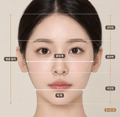
Facial measurements. Labels: facial length; cheekbone width; jaw width; upper, middle and lower face.
The 1:1:1 label illustrates equal vertical thirds as a reference, not an individual treatment target.
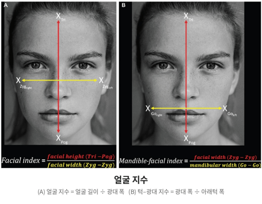
Facial measurement indices. A Facial index: facial height divided by cheekbone width.
B Mandible-facial index: cheekbone width divided by mandibular width.
The anatomical landmarks shown in this figure determine the measurement definition.
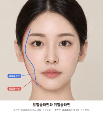
Anterior and outer facial contours. Blue: anterior contour, from the 45-degree cheekbone area toward the jowl.
Red: outer contour, from the lateral cheekbone toward the jaw angle.
Additional anatomical reference images
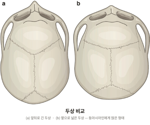
Comparison of head shapes. A A head shape longer from front to back. B A broader head shape.
The original caption describes the broader pattern as more commonly seen in East Asian populations;
individual anatomy varies.
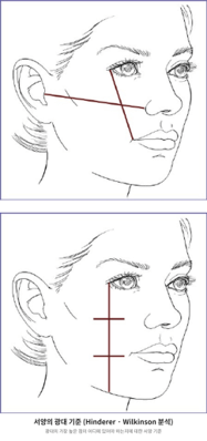
Cheek projection reference lines. The source figure illustrates Hinderer and Wilkinson reference constructions.
These augmentation-oriented references do not by themselves determine a reduction plan.
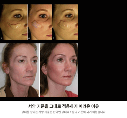
Augmentation examples and reduction planning. Translation of the original caption:
Why Western augmentation guidelines cannot simply be applied to cheekbone reduction in Korean patients.
These are separate reference examples, not the four clinical cases below.
What Are the Risks and Limitations
Cheekbone reduction has potential complications. Published research reports sensory changes, soft-tissue drooping, asymmetry, impaired bone union, restricted mouth opening, bleeding, and facial nerve injury. A meta-analysis of intraoral approaches discusses these outcomes across retrospective studies. Read the complication meta-analysis.
The relevant risk depends on the technique, anatomy, and clinical circumstances. No method can promise an identical outcome for everyone or guarantee that cheek sagging will not occur.
The three-type framework explains differences in planning; it cannot determine suitability, a safe amount of reduction, or the expected result without a clinical assessment.
Clinical Examples How the Planning Priorities Differ
The following summaries come from the supplied clinical article. They illustrate the author’s planning approach and are not independently verified outcome claims. In the original comparisons, before images are on the left and after images are on the right.
- Case 1 Lateral Arch Prominence
The source describes limited resection of the body with substantial inward rotation of the arch.
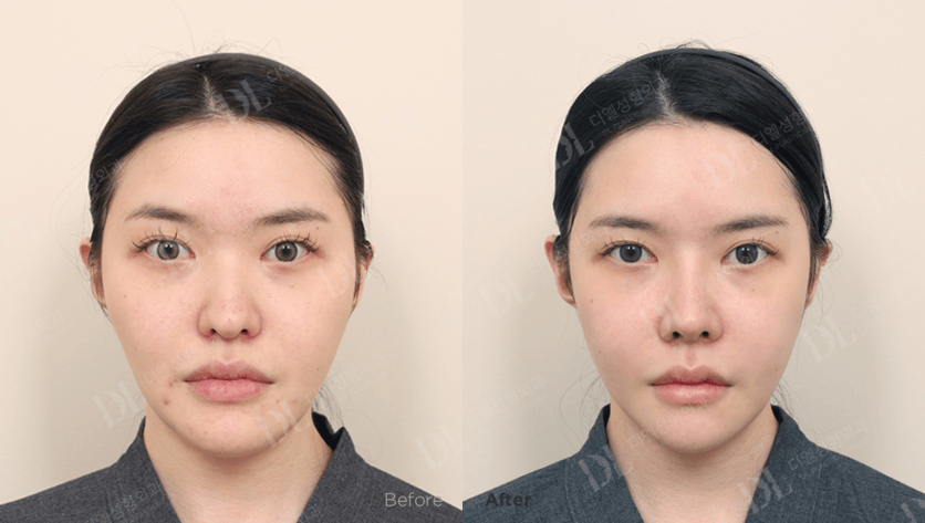
Case 1 lateral arch prominence. Before on the left; after on the right.
The original article describes limited body resection and inward arch rotation.
Other procedures and the postoperative interval are not established here.
- Case 2 45 Degree Prominence
The source describes inward and posterior movement of the body to soften the anterior contour.
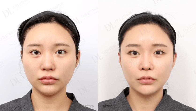
Case 2 45 degree prominence. Before on the left; after on the right.
The source describes inward and posterior body repositioning.
Other procedures and the postoperative interval are not established here.
- Case 3 45 Degree Prominence in a Male Patient
The source describes reducing width while preserving the patient’s desired masculine appearance, with attention to bone thickness, osteotomy placement, and fixation.
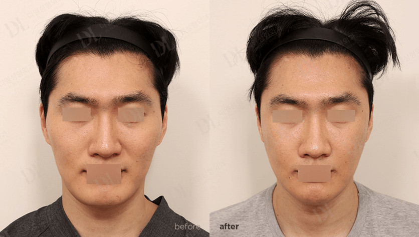
Case 3 45 degree prominence in a male patient. Before on the left; after on the right.
The original privacy masking is preserved.
The source describes width reduction while retaining the desired masculine appearance.
- Case 4 Overall Prominence
The source describes resection and inward rotation while limiting posterior movement because the midface was relatively flat

Case 4 overall prominence. Before on the left; after on the right.
The source describes inward rotation with limited posterior movement.
Other procedures and the postoperative interval are not established here.
What Should You Ask at a Cheekbone Reduction Consultation
1. Is my facial width mainly related to the arch, the cheekbone body, the jaw, or soft tissue?
2. What does the examination or imaging show about each side?
3. Which areas would you reposition, and why?
4. How will you preserve anterior cheek volume and avoid excessive flattening?
5. What bone cuts, resection, and fixation are planned?
6. What are my individual risks, alternatives, and recovery expectations?
7. How will follow-up and assessment of bone healing be arranged?
If you are considering cheekbone reduction in Korea, use the consultation to understand the proposed plan and follow-up arrangements, including care after returning home. An initial discussion can help clarify your questions, but photographs alone do not establish a final surgical plan.
Cheekbone Reduction Surgery Frequently Asked Questions
Is zygoma reduction the same as cheekbone reduction
Yes. “Zygoma reduction” and “reduction malarplasty” are medical terms commonly used for cheekbone reduction surgery. The specific technique can differ, so ask which parts of the cheekbone will be treated.
What is the difference between lateral and 45-degree cheekbone prominence
Lateral prominence mainly affects the arch near the ears and the outer facial width. The term “45-degree prominence” describes a projecting contour in a three-quarter view, often involving the zygomatic body as well as the arch.
Can I identify my cheekbone type from a selfie
A selfie may help explain your concern, but lens distortion, camera angle, lighting, and soft tissue can change the appearance. A clinical assessment, with imaging when appropriate, is needed for surgical planning.
Does every patient need the same amount of bone removed
No. The source article describes different balances of resection, rotation, and repositioning for different patterns. Mixed patterns are also possible. The amount and direction of movement must be individualized.
Can cheekbone reduction make the face look flat
Excessive reduction or unsuitable repositioning may compromise forward cheek projection. Preserving anterior volume is therefore an important planning goal, especially in a relatively flat midface.
Can cheek sagging occur after cheekbone reduction
Yes. Soft-tissue drooping is a reported complication. Surgical planning and fixation require attention, but no technique guarantees that sagging will not occur. Ask the surgeon to explain your individual risk and the proposed approach.
Is cheekbone reduction the same as jaw reduction
No. Cheekbone reduction addresses the zygomatic complex in the midface. Jaw reduction addresses the lower facial skeleton. The need for either procedure depends on which structures contribute to the concern.
How much does cheekbone reduction cost, and how long is recovery
A reliable quote and recovery plan require details of the proposed procedure and the patient’s circumstances. Ask for a written breakdown of fees, planned follow-up, restrictions, and travel timing. This article does not provide a clinic-specific price or recovery timetable.
Discuss Your Cheekbone Shape with a Surgeon
The useful question is not simply “How much can my cheekbones be reduced?” It is “Which structures create my facial width, and how can they be changed while preserving balance?”
Bring front, three-quarter, and side photographs to help explain your concerns, along with details of previous facial procedures. Request an assessment of the cheekbone body, arch, jaw width, and soft tissue before deciding on treatment.

