
Revision Rhinoplasty After Silicone Implant Complications
Can your own rib cartilage replace a silicone nasal implant? In selected cases, a surgeon may use cartilage from your own rib to rebuild the nasal bridge. DL Plastic Surgery describes an approach that uses CT imaging to design a patient-specific mold for finely diced rib cartilage. Whether it is suitable depends on the skin, scar tissue from previous surgery, and the changes needed.
Figure 1 Clinicians with anatomical skull models.
Why Use Diced Rib Cartilage
A surgeon can carve a single piece of rib cartilage to build up the nasal bridge. A solid graft can bend over time, a change called warping. It can also feel firm or shift. Dicing the cartilage avoids having one continuous piece that can bend as a unit and allows small pieces to be used in the graft.
In the technique described here, the cartilage is cut into pieces of about 0.5 mm or smaller. Loose pieces are difficult to shape by hand, so a mold helps define the graft’s length, height, and curve.
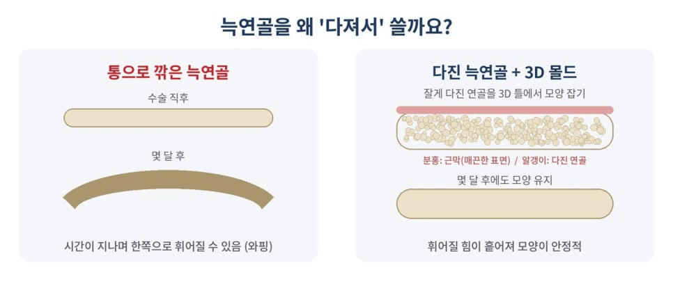
Figure 2 · Why dice rib cartilage? English translation of the labels.
Left · Solid rib cartilage graft
Immediately after surgery; several months later; the graft may bend to one side over time (warping).
Right · Diced rib cartilage + 3D mold
Shape the finely diced cartilage in a 3D mold. Pink layer: fascia (smooth surface).
Particles: diced cartilage.
The lower illustration shows retained shape several months later.
Dicing disperses the forces that can cause warping.
How the Custom Mold Is Designed
DL Plastic Surgery uses CT imaging to design a mold that follows the patient’s nasal anatomy. A standard mold comes in preset sizes; a patient-specific mold can account for the depth of the nasal root between the eyes, the bridge’s curve, and left-to-right asymmetry. The planning concept is similar to that used for a custom silicone implant, but the graft is made from the patient’s own rib cartilage.
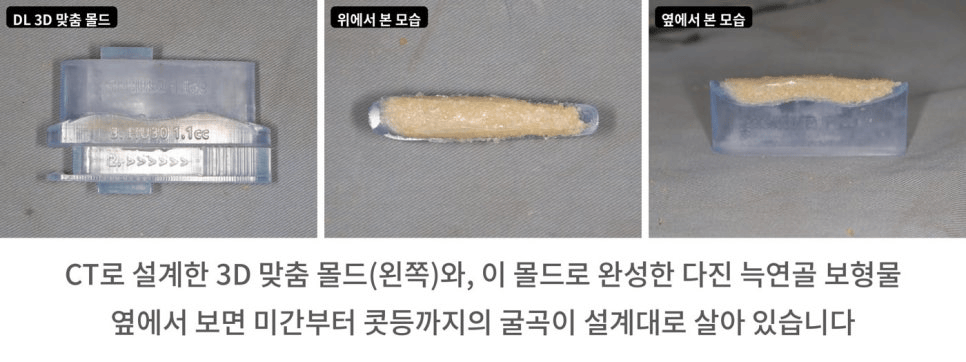
Figure 3 · Left to right
DL 3D Custom Mold · Top View · Side View. The side view shows the planned curve from the nasal root to the bridge.
How the Graft Is Made
The surgeon lines the mold with fascia, a thin layer of connective tissue, and fills it with diced rib cartilage and fibrin glue. The lid compresses the mixture while excess fluid and adhesive drain through side openings. The graft is then removed and transferred to an insertion instrument to help it keep its shape.
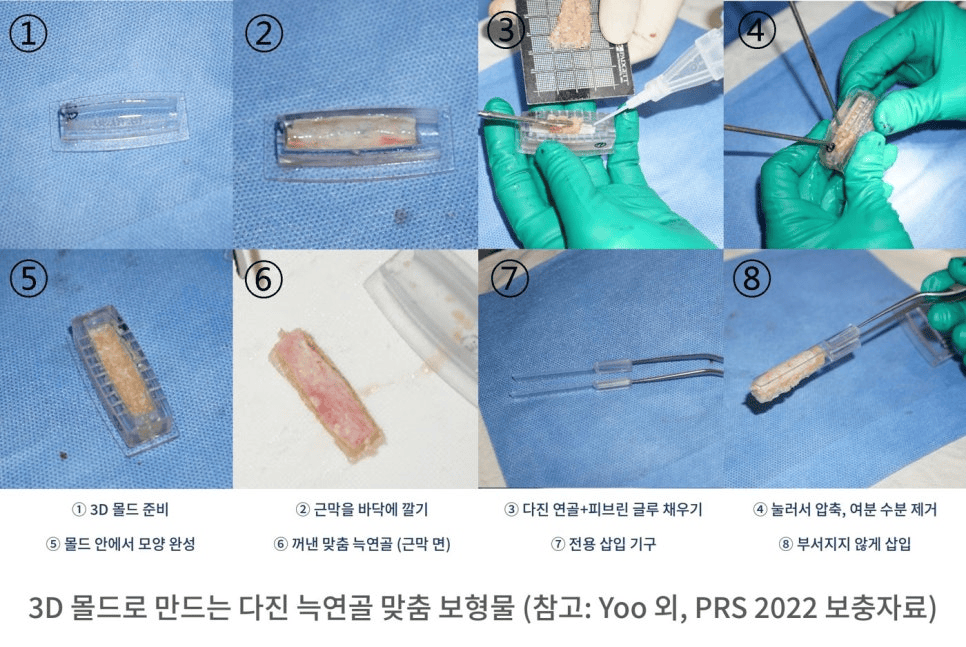
Figure 4 Making a custom diced rib cartilage graft with a 3D mold.
Reference: Yoo et al., Plastic and Reconstructive Surgery, 2022, supplementary material.
1. Prepare the 3D mold.
2. Line the base of the mold with fascia.
3. Add diced cartilage and fibrin glue.
4. Compress the graft and drain excess fluid.
5. Let the graft take shape inside the mold.
6. Remove the custom graft (fascia side shown).
7. Prepare the dedicated insertion instrument.
8. Insert the graft carefully to keep it intact.
Why Add Fascia
The fascia-covered side faces the skin. Fascia can smooth the surface, which matters when thin skin may reveal small irregularities. It also helps hold the diced cartilage together during handling.
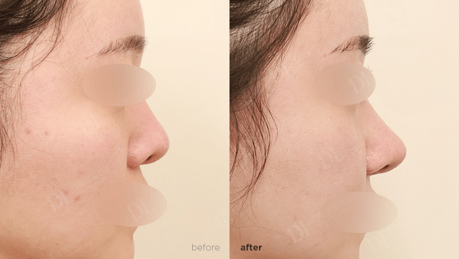
Figure 5 Side profile. Before (left) and after (right).
How the Fit Is Checked Before Insertion
Before inserting the graft, the surgeon can hold it against the bridge to check where it starts near the nasal root, where it ends toward the tip, and how the curve looks from the side. CT helps plan the graft against the underlying anatomy. It cannot fully predict how scar tissue or tight skin will respond, so both need to be assessed in person.
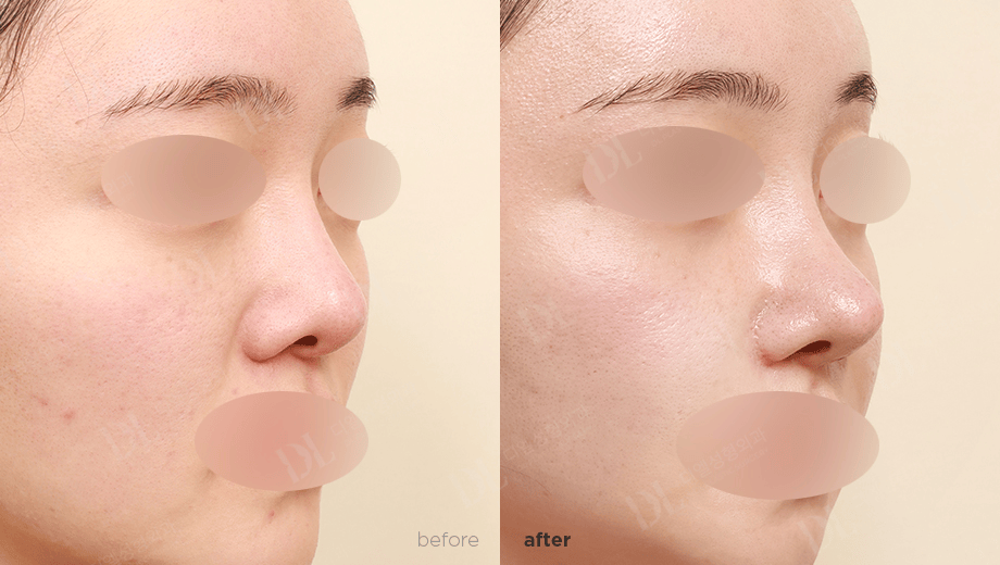
Figure 6 Oblique view. Before (left) and after (right).
What the Research Says About Graft Stability
A 2022 retrospective study by Yoo and colleagues included 80 patients who received a molded, glued diced cartilage graft. Among the 23 patients with 3D scans, bridge height did not decrease significantly between three months and one year. The bridge did not widen from before surgery to one year. In the full group, one patient needed another operation because of graft resorption.
The study evaluated the researchers’ mold. It did not evaluate DL Plastic Surgery’s patient-specific mold or establish how an individual graft will heal. Ask your surgeon about changes in height or contour and whether they recommend taping, splinting, or other follow-up care.
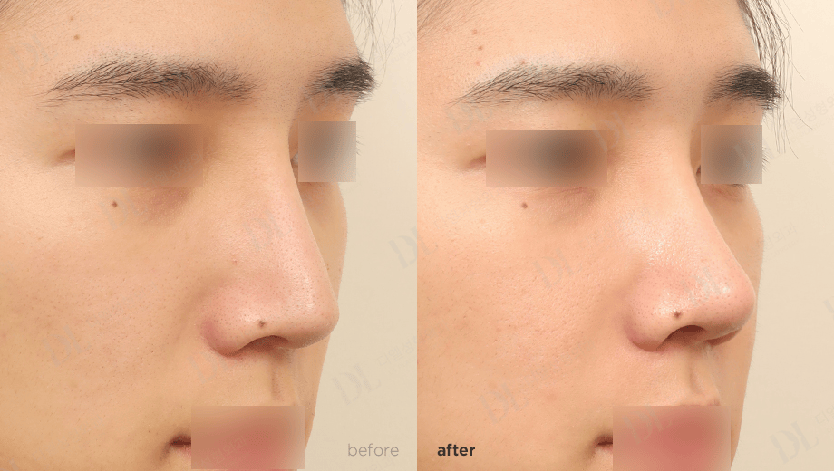
Figure 7 Oblique view. Before (left) and after (right). The clinic watermark in Figures 5–7 reads “DL Plastic Surgery.” Individual results vary.
Frequently Asked Questions
Can diced rib cartilage be used after silicone implant inflammation or contracture?
It may be an option for selected patients. The surgeon first evaluates the previous implant complication, the skin, and how scar tissue affects the shape and mobility of the nose.
How is a patient-specific mold different from a standard mold?
A standard mold comes in set sizes. A patient-specific mold is designed from a CT scan to reflect the planned bridge height, curve, and asymmetry.
Can a diced rib cartilage graft lose height over time?
Yes. The body can absorb some of the cartilage, which may change bridge height or contour. One-year study results cannot predict an individual outcome.
Questions to Ask at Your Consultation
- How do my previous implant complications, scars, and skin condition affect the plan?
- What does my CT scan show about the nasal bones, nasal root, and asymmetry?
- Will you use a standard mold or one designed from my CT scan?
- Will you use fascia on the side of the graft that faces the skin?
- What changes might I see during healing, and what follow-up will I need?
Research Reference
Yoo SH, Kim DH, Jang YJ. Dorsal Augmentation Using a Glued Diced Cartilage Graft Fashioned with a Newly Developed Mold in Asian Rhinoplasty. Plastic and Reconstructive Surgery. 2022;150(4):757e–766e. doi:10.1097/PRS.0000000000009568.

